Research
Overview
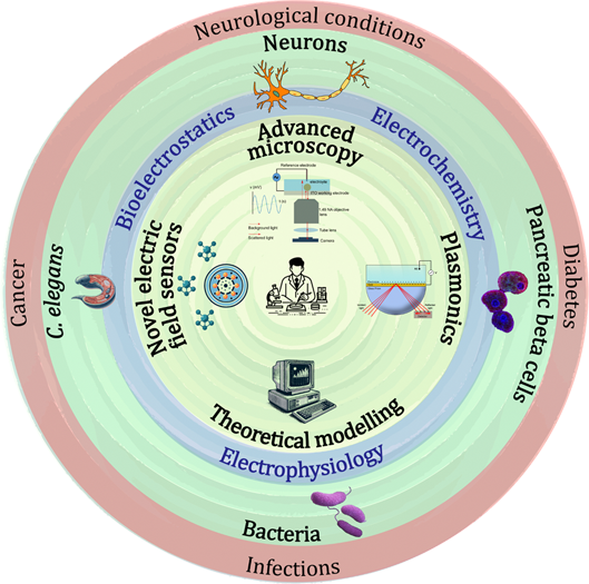
We conduct interdisciplinary research that explores nanoscale electric and electrostatic charge with a particular focus on living systems in healthy and diseased conditions.
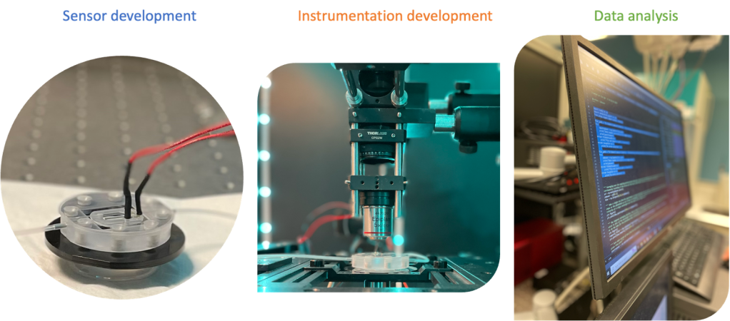
We develop novel methods from initial sensing concepts to demonstrating innovative applications in biology, electrochemistry, and materials. This process involves sensor development using the state-of-the-art nano/micro fabrication technology. To achieve this, we exploit various charge sensitive optical phenomena including surface plasmonics. Our lab builds precision optical measurement systems along with customised data analysis techniques to maximise information extraction. Additionally, we conduct theoretical research that integrates physical chemistry models of electrified media with various optical models to understand charge carriers and light interactions. Our methods are demonstrated using living system models spanning the range of a single cell to a whole living organism.
3D electrostatic charge mapping in living systems
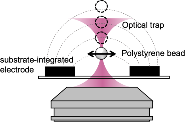
Understanding diseases such as cancer requires a deep understanding of molecular and cellular functions. Hence, there is a pressing demand for tools to explore the characteristics of microscopic living systems, including often-overlooked electrostatic charge, which influences biochemical reactions and cellular behaviours. Current techniques for studying electrostatic charge have limitations, particularly with complex 3D biological samples. Our research aims to uncover electrostatic properties developing a new capability to detect tiny charges and forces within microenvironments. Collaborating with Prof. Amanda Wright, we combine optical trapping with custom electrochemical setups to create a unique microscopy technique for mapping changing electrostatic charges around live biological samples in 3D, opening new avenues for understanding electrostatic charge in cancers.
Novel deciphering of disordered neural circuits in living organisms
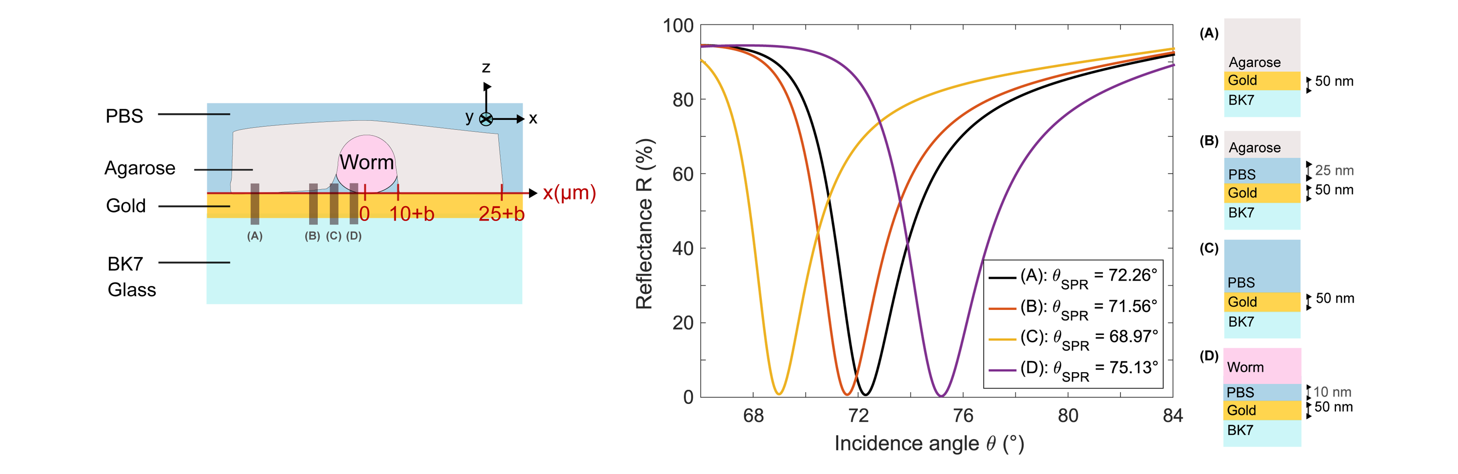
The brain and nervous system, complex in both humans and mammals, consist of billions of interconnected neurons forming dense structures. Understanding these systems, especially in neurological disorders, challenges neuroscience. Caenorhabditis elegans is widely used as an in vivo model due to its transparent anatomy, fully sequenced genome, and short lifespan. Its well-understood nervous system enables detailed studies on behaviour-influencing neural circuits. However, current tools have limited ability to track electrical signals in living organisms. Our lab develops a new label-free imaging technique for bioelectrical signalling in whole living organisms, starting with C. elegans. This innovative approach aims to provide a novel approach to understanding and addressing neurological conditions using an in vivo model of whole living organisms.
Electrical Impedance Microscopy
Living organisms can be partly considered electrical systems, as they generate and conduct electrical signals to coordinate various functions, such as cognition, muscle contraction, and hormone release. However, due to the absence of suitable measurement tools, bioelectrical properties remain poorly understood. This research theme is directed at developing a novel microscopy technology platform for studying the electrical properties of biological tissue with sub-microscopic resolution. Our vision is to create a multimodal impedance optical microscope to generate fine-grained images of the electrical properties of single cells in health and disease.
Label-free imaging of pancreatic beta cell networks in diabetes
The World Health Organization reports that around 422 million people worldwide are affected by diabetes, with approximately 1.5 million deaths attributed to the disease each year. Interdisciplinary research endeavours are underway to understand and address diabetes, including efforts to develop new diagnostic, management, and treatment methods. Our lab is actively contributing to this research by developing a novel label-free optical method for functional imaging of insulin-secreting beta cells. Our goal is to provide the diabetes research community with a new tool for investigating beta cell networks in both healthy and diabetic conditions.
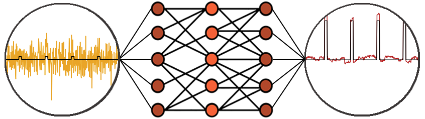
Ultrasensitive voltage detection using AI
Modern microscopic techniques, including our new impedance, voltage, and electrostatic microscopes, face the challenge of extracting small signals from highly noisy environments. In response, our lab is leveraging the potential of artificial intelligence methods to exceed the capabilities of traditional signal processing techniques. Our approach aims to provide novel methods for detecting minute signals at the microscopic scale, enabling a deeper understanding of the samples under investigation.
Label-free optical voltage sensing
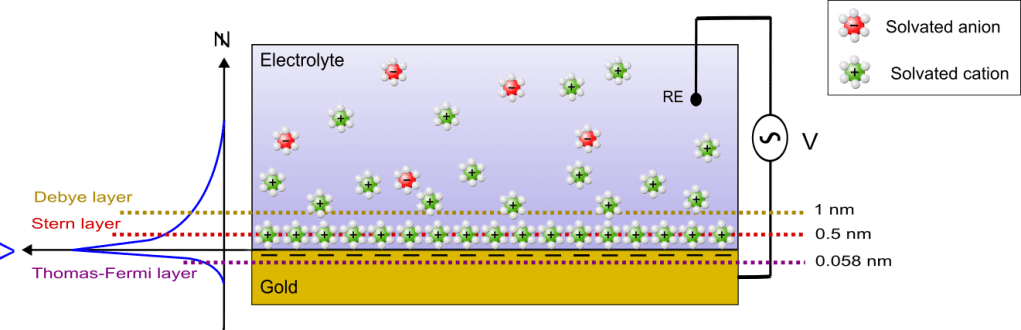
This direction aims to provide ways for highly sensitive detection of changes in voltage without the need for dyes or scanning probes. We have previously employed techniques such as surface plasmon resonance (SPR) to measure small voltage variations. We investigate new techniques for enhanced and widely applicable optical label-free voltage sensing, for instance using thin metallic films with various thicknesses combined with a wide range of optical configurations.